Julia Crim: Imaging Anatomy: Musculoskeletal, Gebunden
Imaging Anatomy: Musculoskeletal
Buch
Derzeit nicht erhältlich.
Lassen Sie sich über unseren eCourier benachrichtigen, falls das Produkt bestellt werden kann.
Lassen Sie sich über unseren eCourier benachrichtigen, falls das Produkt bestellt werden kann.
- Verlag:
- Elsevier LTD, 06/2026
- Einband:
- Gebunden
- Sprache:
- Englisch
- ISBN-13:
- 9780443433450
- Umfang:
- 816 Seiten
- Nummer der Auflage:
- 26003
- Ausgabe:
- 3rd edition
- Gewicht:
- 450 g
- Erscheinungstermin:
- 10.6.2026
- Hinweis
-
Achtung: Artikel ist nicht in deutscher Sprache!
Klappentext
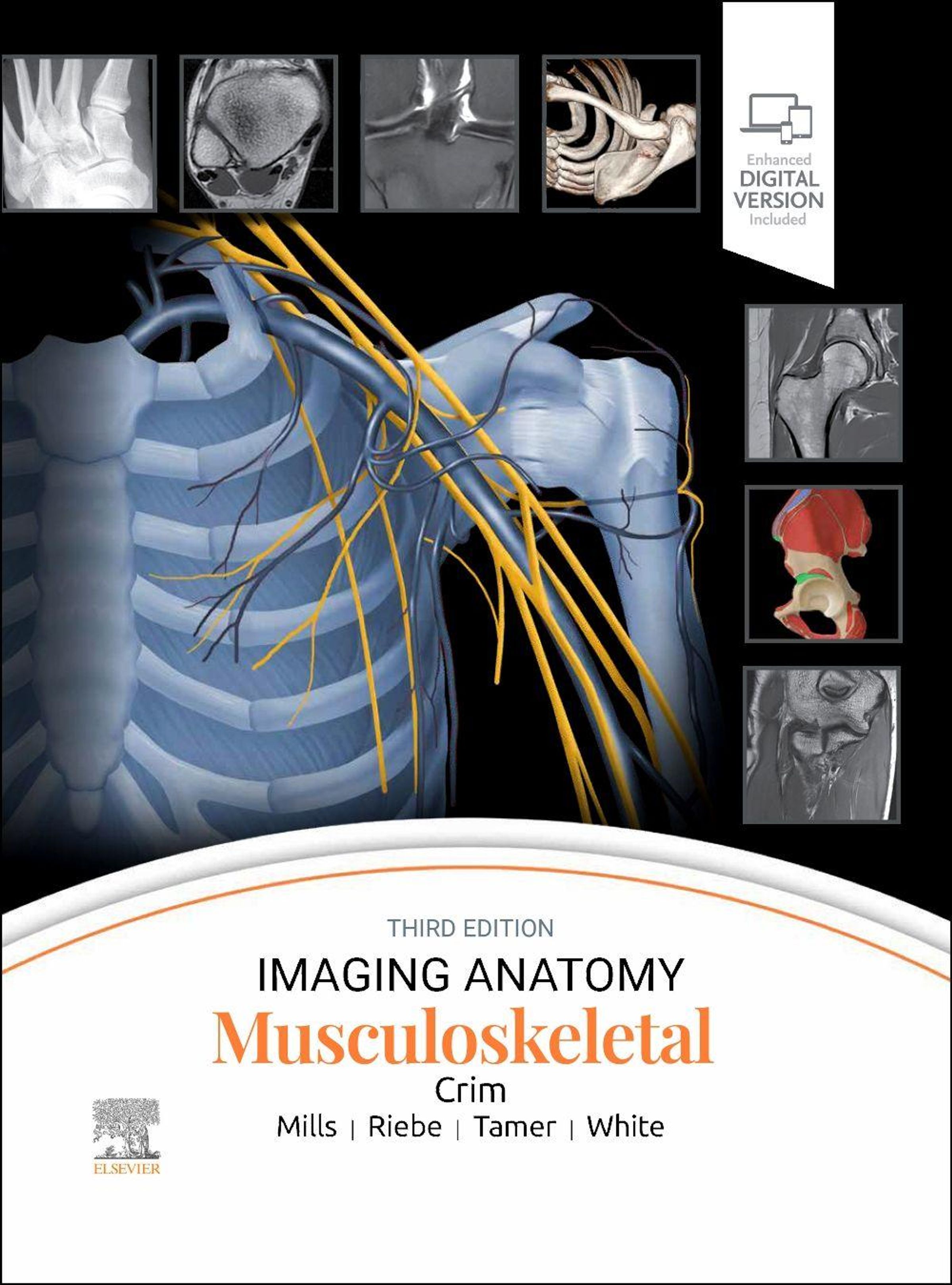
The third edition of Imaging Anatomy: Musculoskeletal is a must-have reference for your practice. New chapters, updated content, and all-new 3T and 7T MR images provide detailed insight into both human anatomy and imaging pitfalls. Dr. Julia Crim and her team of specialists present an easy-to-read, complete anatomic atlas of the musculoskeletal system that features more than 1, 900 high-quality images and illustrations as well as carefully chosen references for further reading. Imaging Anatomy: Musculoskeletal is a highly visual and accessible resource for radiologists at all levels of experience, as well as orthopedic surgeons, sports medicine practitioners, and all others involved with musculoskeletal imaging.
- Features all-new 3T and 7T MR images that provide exceptional imaging detail
- Illustrates pediatric skeletal development
- Presents radiographic, CT, and MR images complemented bylarge, extensively labeled artists' illustrations throughout
- Includes new chapters on the brachial plexus, scapula, clavicle, and sternum that reflect the enlarging scope of musculoskeletal practice as well as new introductory chapterselucidating bone maturation, bone marrow, and the musculotendinous unit
- Features overview chapters for each region that provide essential information, radiographs, and expert medical illustrations as well as focused chapters that dive deeper into the most complex anatomic issues, such as posterolateral corner of the knee
- Provides detailed anatomy in at least 3 planes in MR atlas chapters
- Discusses common normal variants, imaging pitfalls, and clinical applications for each area
- Contains updated text to reflect improved MRvisualization of small structures and current clinical concerns related to anatomy
- Employs a time-saving, bulleted format that distills complex information for easy comprehension
- Includes a complimentary eBook with access to everything in the print version as well as additional images, text, and references, with the ability to search, customize your content, and have content read aloud